Poster vincitore nella sezione ricerca di base al congresso nazionale SIPMO
del 15/06/2011 (Pugnochiuso – Fg)

Domenico Tesi, Giuseppe Ficarra
Oral linear epidermal nevus: a review of the literature and report of two new cases
Head Neck Pathol 2010 Jun;4(2):139-43.
doi: 10.1007/s12105-010-0165-7. Epub 2010 Feb 9.
Abstract
Linear epidermal nevus (LEN) is a sporadic hamartomatous lesion of the skin due to the proliferation
of clones of embryonic ectodermal cells, which are arranged according to a typical linear configuration
known as Blaschko’s lines. Oral involvement of LEN is very rare and few cases have been reported
in the medical literature. We report two new cases of LEN with exclusive oral involvement, which
presented with the typical unilateral or midline distribution. Oral LEN presents as an exophytic
lesion with well defined borders and a verrucous or papillary surface which correspond,
histologically, to epithelial papillomatosis with a moderate degree of hyperkeratosis and acanthosis.
Oral LEN appears mainly at birth, grows slowly during childhood and stabilize by adolescence.
Localized lesions do not show any recurrence after surgical removal but widespread lesions seem
to have more risk of recurrence and are more difficult to manage. Functional problems and
malignant transformation have not been reported.

Singh M, Ficarra G, Tesi D, Massi D, Stefanato CM
A solitary ulcer of the tongue: a quiz. Primary syphilitic chancre of the tongue
Acta Derm Venereol. 2014 Mar;94(2):254-5. doi: 10.2340/00015555-1655.PMID: 23824304

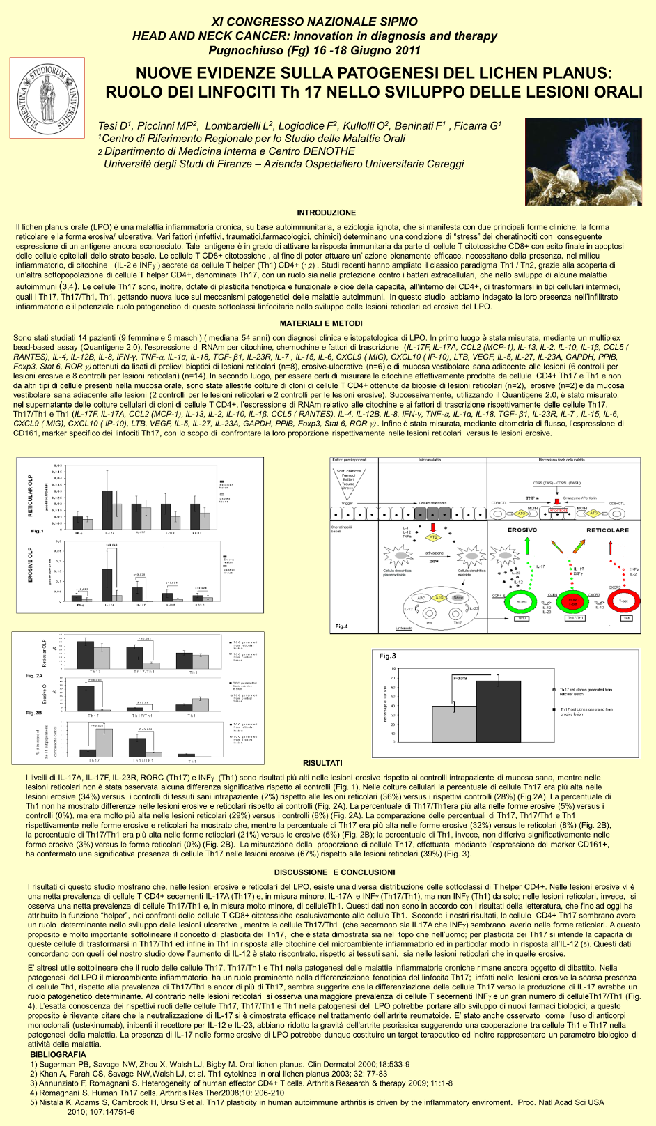
M-P Piccinni 1, L Lombardelli, F Logiodice, D Tesi, O Kullolli, R Biagiotti, Mg Giudizi,
S Romagnani, E Maggi, G Ficarra
Potential pathogenetic role of Th17, Th0, and Th2 cells in erosive and reticular
oral lichen planus
Oral Dis
. 2014 Mar;20(2):212-8.
doi: 10.1111/odi.12094. Epub 2013 Apr 5.
Abstract
Objectives: The role of Th17 cells and associated cytokines was investigated in oral lichen planus.
Material and methods: 14 consecutive patients with oral lichen planus were investigated. For biological studies,
tissues were taken from reticular or erosive lesions and from normal oral mucosa (controls) of the same patient.
mRNA expression for IL-17F, IL-17A, MCP-1, IL-13, IL-2, IL-10, IL-1β, RANTES, IL-4, IL-12B, IL-8, IFN-γ, TNF-α, IL-1α,
IL-18, TGF-β1, IL-23R, IL-7, IL-15, IL-6, MIG, IP-10, LTB, VEGF, IL-5, IL-27, IL-23A, GAPDH, PPIB, Foxp3, GATA3, and
RORC was measured using the QuantiGene 2.0.
Results: Results showed that Th17-type and Th0-type molecules’ mRNAs, when compared with results obtained
from tissue controls, were increased in biopsies of erosive lesions, whereas Th2-type molecules’ mRNAs were
increased in reticular lesions. When the CD4+ T-cell clones, derived from oral lichen planus tissues and tissue
controls, were analyzed, a higher prevalence of Th17 (confirmed by an increased CD161 expression) and Th0 CD4+ T
clones was found in erosive lesions, whereas a prevalence of Th2 clones was observed in reticular lesions.
Conclusions: Our data suggest that Th17, Th0, and Th2 cells, respectively, may have a role in the pathogenesis
of erosive and reticular oral lichen planus.
Keywords: Th17; autoimmunity; lichen planus; lymphocytes; mucosa; oral.
© 2013 John Wiley & Sons A/S. Published by John Wiley & Sons Ltd.
https://pubmed.ncbi.nlm.nih.gov/23556506/